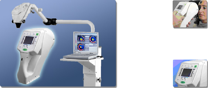
Computer assisted Corneal Topography

Corneal topography is a process for mapping the surface curvature of the cornea, similar to making a contour map of land. The cornea is a clear membrane that covers the front of the eye and is responsible for about 70 percent of the eye’s focusing power. To a large extent, the shape of the cornea determines the visual ability of an otherwise healthy eye. A perfect eye has an evenly rounded cornea, but if the cornea is too flat, too steep, or unevenly curved, less than perfect vision results.
The purpose of corneal topography is to produce a detailed description of the shape and power of the cornea. Using computerized imaging technology, the 3-dimensional map produced by the corneal topographer aids an ophthalmologist in the diagnosis, monitoring, and treatment of various visual conditions.
Corneal topography is not a routine test. Rather, it is used in diagnosing certain types of problems, in evaluating a disease’s progression, in fitting some types of contact lenses, and in planning surgery. It is commonly used in preparing for refractive eye surgery. The corneal topography map is used in conjunction with other tests to determine exactly how much corneal tissue will be removed to correct the visual defect.Corneal topography is used in the diagnosis and management of various corneal curvature abnormalities and diseases such as:• Keratoconus, a degenerative condition that causes a thinning of the cornea
- Corneal transplants
- Corneal scars or opacities
- Corneal deformities
- Fitting contact lenses
- Irregular astigmatism following corneal transplantation
- Planning refractive surgery
- Postoperative cataract extraction with acquired astigmatism
